The beginning of a new organism is given by a fertilized egg (with the exception of cases of parthenogenesis and vegetative reproduction). Fertilization is the process of fusion of two germ cells (gametes) with each other, during which two different functions are carried out: sexual (combining the genes of two parental individuals) and reproductive (the emergence of a new organism). The first of these functions includes the transfer of genes from parents to offspring, the second - the initiation in the cytoplasm of the egg of those reactions and movements that allow further development. As a result of fertilization, a double (2p) set of chromosomes is restored in the egg. The centrosome, introduced by the sperm, after doubling forms a fission spindle, and the zygote enters the 1st stage of embryogenesis - the stage of crushing. As a result of mitosis, 2 daughter cells - blastomeres - are formed from the zygote.
Prezygotic period
The prezygotic period of development is associated with the formation of gametes (gametogenesis). The formation of oocytes begins in women even before they are born and is completed for each given oocyte only after its fertilization. By the time of birth, the female fetus in the ovaries contains about two million first-order oocytes (these are still diploid cells), and only 350 - 450 of them will reach the stage of second-order oocytes (haploid cells), turning into eggs (one at a time during one menstrual cycle). Unlike women, germ cells in the testes (testicles) in men begin to form only with the onset of puberty. The duration of the period of sperm formation is approximately 70 days; for one gram of testicle weight, the number of spermatozoa is about 100 million per day.
Fertilization
Fertilization - the fusion of a male reproductive cell (sperm) with a female (egg, ovum), leading to the formation of a zygote - a new unicellular organism. The biological meaning of fertilization is the unification of the nuclear material of male and female gametes, which leads to the unification of paternal and maternal genes, the restoration of the diploid set of chromosomes, as well as the activation of the egg, that is, its stimulation for embryonic development. The connection of the egg with the sperm usually occurs in the funnel-shaped part of the fallopian tube during the first 12 hours after ovulation.
The seminal fluid, entering the woman's vagina during sexual intercourse, usually contains from 60 to 150 million spermatozoa, which, thanks to movements at a speed of 2-3 mm per minute, constant undulating contractions of the uterus and tubes and an alkaline environment, already after 1-2 minutes after intercourse, they reach the uterus, and after 2-3 hours - the end sections of the fallopian tubes, where they usually merge with the egg. There are monospermic (one sperm enters the egg) and polysperm (two or more sperm enter the egg, but only one sperm nucleus fuses with the egg nucleus) fertilization. Preservation of sperm activity during their passage in the genital tract of a woman is facilitated by the slightly alkaline environment of the cervical canal of the uterus, filled with a mucous plug. During orgasm during sexual intercourse, the mucous plug from the cervical canal is partially pushed out, and then retracted into it and thereby contributes to a faster entry of spermatozoa from the vagina (where it is normal for healthy woman slightly acidic environment) to a more favorable environment of the cervix and uterine cavity. The passage of spermatozoa through the mucous plug of the cervical canal is also facilitated by the sharp increase in mucus permeability on the days of ovulation. On the remaining days of the menstrual cycle, the mucous plug has a significantly lower permeability for spermatozoa.
Many spermatozoa located in the genital tract of a woman can retain the ability to fertilize for 48-72 hours (sometimes even up to 4-5 days). An ovulated egg remains viable for approximately 24 hours. Given this, the most favorable time for fertilization is the period of rupture of a mature follicle, followed by the birth of an egg, as well as the 2-3rd day after ovulation. Women using a physiological method of contraception should be aware that the timing of ovulation may fluctuate, and the viability of the egg and sperm can be significantly longer. Shortly after fertilization, zygote cleavage and embryo formation begin.
ZygoteZygote (Greek zygote paired) is a diploid (containing a complete double set of chromosomes) cell resulting from fertilization (the fusion of an egg and a sperm cell). The zygote is a totipotent (that is, capable of producing any other) cell. The term was introduced by the German botanist E. Strasburger.
In humans, the first mitotic division of the zygote occurs approximately 30 hours after fertilization, which is due to the complex processes of preparation for the first act of crushing. Cells formed as a result of crushing the zygote are called blastomeres. The first divisions of the zygote are called "crushing" because the cell is crushed: after each division, the daughter cells become smaller and smaller, and there is no stage of cell growth between divisions.
Development of the zygote The zygote either begins to develop immediately after fertilization, or is dressed in a dense shell and for some time turns into a resting spore (often called a zygospore) - typical of many fungi and algae.
Splitting upPeriod embryonic development multicellular animal begins with the crushing of the zygote and ends with the birth of a new individual. The cleavage process consists in a series of successive mitotic divisions of the zygote. The two cells formed as a result of a new division of the zygote and all subsequent generations of cells at this stage are called blastomeres. During crushing, one division follows another, and the resulting blastomeres do not grow, as a result of which each new generation of blastomeres is represented by smaller cells. This feature of cell divisions during the development of a fertilized egg determined the appearance of a figurative term - crushing of the zygote.
At different types Animal eggs differ in the quantity and nature of the distribution of reserve nutrients (yolk) in the cytoplasm. This largely determines the nature of the subsequent fragmentation of the zygote. With a small amount and uniform distribution of the yolk in the cytoplasm, the entire mass of the zygote divides with the formation of identical blastomeres - complete uniform crushing (for example, in mammals). When the yolk accumulates predominantly at one of the poles of the zygote, uneven fragmentation occurs - blastomeres are formed that differ in size: larger macromeres and micromeres (for example, in amphibians). If the egg is very rich in yolk, then its part, free of yolk, is crushed. So, in reptiles, birds, only the disc-shaped section of the zygote at one of the poles, where the nucleus is located, undergoes crushing - incomplete, discoidal crushing. Finally, in insects, only the surface layer of the cytoplasm of the zygote is involved in the crushing process - incomplete, superficial crushing.
As a result of crushing (when the number of dividing blastomeres reaches a significant number), a blastula is formed. In a typical case (for example, in the lancelet), the blastula is a hollow ball, the wall of which is formed by a single layer of cells (blastoderm). The blastula cavity - blastocoel, otherwise called the primary body cavity, is filled with fluid. In amphibians, the blastula has a very small cavity, and in some animals (such as arthropods), the blastocoel may be completely absent.
gastrulationAt the next stage of the embryonic period, the process of gastrula formation takes place - gastrulation. In many animals, the formation of the gastrula occurs by invagination, i.e. protrusions of the blastoderm at one of the blastula poles (with intensive reproduction of cells in this zone). As a result, a two-layer, bowl-shaped embryo is formed. The outer layer of cells is the ectoderm, and the inner layer is the endoderm. The internal cavity that occurs when the wall of the blastula protrudes, the primary intestine, communicates with the external environment through an opening - the primary mouth (blastopore). There are other types of gastrulation. For example, in some coelenterates, the endoderm of the gastrula is formed by immigration, i.e. "eviction" of a part of blastoderm cells into the cavity of the blastula and their subsequent reproduction. The primary mouth is formed by rupture of the wall of the gastrula. With uneven crushing (in some worms, mollusks), the gastrula is formed as a result of fouling of macromeres with micromeres and the formation of endoderm due to the first. Often different methods of gastrulation are combined.
In all animals (except for sponges and coelenterates - bilayer animals), the gastrulation stage ends with the formation of another layer of cells - the mesoderm. This "cell layer is formed between the ento- and ectoderm. There are two known ways of laying the mesoderm. In annelids, for example, two large cells (teloblasts) are isolated in the blastopore region of the gastrula. Reproducing, they give rise to two mesodermal stripes, of which (partly due to divergence of cells, partly as a result of the destruction of part of the cells inside the mesodermal strips), coelomic sacs are formed - the teloblastic method of laying the mesoderm.In the enterocelous method (echinoderms, lancelet, vertebrates), as a result of protrusion of the wall of the primary intestine, lateral pockets are formed, which then separate and become coelomic In both cases of mesoderm initiation, coelomic sacs grow and fill the primary body cavity. The mesodermal layer of cells lining the inside of the body cavity forms the peritoneal epithelium. The cavity that thus replaced the primary is called the secondary body cavity, or coelom. In the case of the teloblastic method of mesoderm initiation The blastopore develops into the mouth opening of an adult animal. Such organisms are called protostomes. In deuterostomes (with the enterocoelous method of laying the mesoderm), the blastopore overgrows or turns into an anus, and the mouth of an adult occurs a second time, by protrusion of the ectoderm.
The formation of three germ layers (ecto-, ento- and mesoderm) completes the stage of gastrulation, and from this moment the processes of histo- and organogenesis begin. As a result of cell differentiation of the three germ layers, various tissues and organs of the developing organism are formed. At the end of the last century (largely thanks to the studies of I. I. Mechnikov and A. O. Kovalevsky) it was established that in different animal species the same germ layers give rise to the same organs and tissues. From the ectoderm, the epidermis with all derivative structures and the nervous system are formed. Due to the endoderm, the digestive tract and associated organs (liver, pancreas, lungs, etc.) are formed. The mesoderm forms the skeleton, vascular system, excretory apparatus, gonads. Although today the germ layers are not considered strictly specialized, nevertheless, their homology in the vast majority of animal species is obvious, which indicates the unity of the origin of the animal kingdom.
During the embryonic period, there is an increase in the rate of growth and differentiation in developing organisms. If growth does not occur during cleavage and the blastula (in terms of mass) can be significantly inferior to the zygote, then, starting from the process of gastrulation, the mass of the embryo rapidly increases (due to intensive cell reproduction). The processes of cell differentiation begin at the earliest stage of embryogenesis - crushing and underlie primary tissue differentiation - the emergence of three germ layers (embryonic tissues). Further development of the embryo is accompanied by an ever-increasing process of differentiation of tissues and organs. As a result of the embryonic period of development, an organism is formed that is capable of independent (more or less) existence in the external environment. A new individual is born either as a result of hatching from an egg (in oviparous animals) or exit from the mother's body (in viviparous).
Histo - and organogenesisHisto - and organogenesis of the embryo are carried out as a result of reproduction, migration, differentiation of cells, its components, the establishment of intercellular contacts and the death of some cells. The 317th to the 20th day continues the presomitic period from the 20th day the somite period of development begins. On the 20th day of embryogenesis, through the formation of trunk folds (cephalocaudal and lateral), the embryo itself is separated from extraembryonic organs, as well as its flat shape is changed to a cylindrical one. At the same time, the dorsal parts of the mesoderm of the embryo are divided into separate segments located on both sides of the chord - somites. On the 21st day, there are 2-3 pairs of somites in the body of the embryo. Somites begin to form from the III pair, I and II pairs appear somewhat later. The number of somites gradually increases: on the 23rd day of development there are 10 pairs of somites, on the 25th - 14 pairs, on the 27th - 25 pairs, at the end of the fifth week the number of somites in the embryo reaches 43-44 pairs. Based on the calculation of the number of somites, it is possible to approximately determine the timing of development (somitic age) of the embryo.
From the outer part of each somite, a dermatome arises, from the inner - a sclerot, from the middle - a myot. The dermatome becomes the source of the skin dermis, the sclerote becomes the source of cartilage and bone tissue, and the myotome becomes the source of the skeletal muscles of the dorsal part of the embryo. The ventral sections of the mesoderm - splanchnotome - are not segmented, but are divided into visceral and parietal sheets, from which serous membranes of internal organs develop, muscle heart and adrenal cortex. Blood vessels, blood cells, connective and smooth muscle tissue of the embryo are formed from the mesenchyme of the splanchnotome. The section of the mesoderm that connects the somites with the splanchnotome is divided into segmented legs - the nephrogonoth, which serve as a source for the development of the kidneys and gonads, as well as the paramesonephric ducts. Of the latter, the epithelium of the uterus and oviduct is formed.
In the process of differentiation of the germinal ectoderm, the neural tube, neural crests, placodes, skin ectoderm and prechordal plate are formed. The process of neural tube formation is called neurulation. It consists in the formation of a slit-like depression on the surface of the ectoderm; the thickened edges of this depression (neural folds) fuse to form the neural tube. Brain vesicles form from the cranial part of the neural tube, which is the rudiment of the brain. On both sides of the neural tube (between the latter and the skin ectoderm), groups of cells are separated, from which neural crests are formed. Neural crest cells are capable of migrating. Cells migrating in the direction of the dermatome give rise to pigment cells - melanocytes; neural crest cells that migrate towards the abdominal cavity give rise to the sympathetic and parasympathetic ganglia, the adrenal medulla. From the cells of the neural crests, which did not migrate, ganglionic plates are formed, from which the spinal and peripheral autonomic nerve ganglia develop. The ganglia of the head and the nerve cells of the organ of hearing and balance are formed from the placodes.
Splitting up(segmentation) in individual representatives of the category of vertebrates generally has the same course; however, as already mentioned above, it was influenced by factors that during phylogenesis influenced development in the form of consequences of the influence of the internal and external environment in which organisms lived during their ancestral development (cenogenetic factors).
When observing changes, occurring in eggs according to the phylogenetic development of eggs of individual representatives of the vertebrate category, it can be seen that the egg cells differ significantly from each other in the content of the nutrient and building substance - the yolk. The egg cells of the lancelet (Amfioxus), an organism that is phylogenetically considered the lowest organized creature, but which already has a strong dorsal region, are among the oligolecital.
However, in accordance with phylogenetic development, the amount of yolk in the eggs of vertebrates, which are phylogenetically the most highly organized organisms, increases more and more, reaching a maximum amount in avian eggs, which are relatively very large and polylecital. Under the influence of coenogenetic factors (factors influencing from the external environment and determined by changes in lifestyle and, consequently, development), the amount of yolk in the process of phylogenetic development towards humans decreases more and more, due to which the eggs of humans and higher mammals become again (secondarily) oligolecithal .
Having a variable amount yolk has, as mentioned above, a significant impact on the process of crushing the egg. Egg cells with a low yolk content (oligolecital) are completely crushed, that is, the entire substance of a fertilized egg is divided into new cells, blastomeres (holoblastic type eggs) during crushing. On the contrary, in eggs containing more yolk, or even a large amount of yolk (polylecital), cleavage furrows continuously crush only a smaller part of the ooplasm, located at the so-called animal pole, where there are fewer yolk granules (meroblastic type eggs).
In accordance with this, individual representatives of the category vertebrates, the following types of crushing are distinguished.
1. Complete crushing. Complete, total crushing includes those cases when, in the process of crushing division, the entire fertilized egg cell is divided and crushing furrows spread over its entire surface. According to this type, egg cells of the holoblastic species are crushed. Depending on the content of a greater or lesser amount of yolk in the ooplasm, as well as depending on its distribution in the ooplasm, during crushing, blastomeres of either relatively the same size (complete uniform, equal, or adequal cleavage) or blastomeres of various sizes, namely larger in the area with a high content of yolk and smaller in the place where the yolk is less (complete uneven, inequal crushing). Larger blastomeres are called macromeres, smaller ones are called micromeres.
Full equal, or adequal, crushing is characteristic of oligolecithal, isolecithal eggs (lancelet, higher mammals and humans); according to the complete inequal type, mesolecithal egg cells of an anisolecital and moderately telolecithal species (some lower fish and amphibians) are crushed.
2. Partial, partial, crushing. By partial type, egg cells are crushed, containing a significant amount of yolk (polylecithal eggs), which, due to their large sizes cleavage furrows during cell division penetrate only into the region of the animal pole, where the cell nucleus is located and where the ooplasm layer contains fewer yolk granules (higher fish, reptiles, birds and some lower mammals, oviparous).
With such crushing at the animal pole of a relatively large egg, only a round field (disk) is crushed, while the remainder of the egg cell (yolk ball) remains unshattered (partial disc-shaped crushing). In insects, their polylecital centrolecithal oocytes, although they are crushed over the entire surface, but the center of the cell, containing a large amount of yolk, remains not crushed (partial surface crushing).
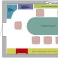
In the above figure individual types of egg cells are shown depending on the content and distribution of the yolk in the ooplasm, as well as depending on the corresponding type of crushing.
Embryogenesis (Greek embryon - embryo, genesis - development) - early period individual development organism from the moment of fertilization (conception) to birth, is initial stage ontogenesis (Greek ontos - being, genesis - development), the process of individual development of the organism from conception to death.
The development of any organism begins as a result of the fusion of two sex cells (gametes), male and female. All cells of the body, despite differences in structure and functions, are united by one thing - a single genetic information stored in the nucleus of each cell, a single double set of chromosomes (except for highly specialized blood cells - erythrocytes, which do not have a nucleus). That is, all somatic (soma - body) cells are diploid and contain a double set of chromosomes - 2 n, and only germ cells (gametes) that form in specialized sex glands (testes and ovaries) contain a single set of chromosomes - 1 n.
When the sex cells merge, a cell is formed - a zygote, in which a double set of chromosomes is restored. Recall that the nucleus of a human cell contains 46 chromosomes, respectively, germ cells have 23 chromosomes.
The sex of the child is determined by the ratio of chromosomes during fertilization. If the egg is fertilized by a sperm with an X sex chromosome, then two x chromosomes are formed in the zygote ( female body). When a spermatozoon with a y chromosome is fertilized, a combination of XY (male body) is formed in the zygote. Since meiosis produces the same number of sperm with X and Y chromosomes, theoretically the number of newborn boys and girls should be the same, but in reality, 103 girls are born for every 100 boys. This is due to the greater sensitivity of male fetuses to various adverse and damaging factors.
Embryogenesis types

The type of embryogenesis is a set of features that provide a developing organism with a connection with the environment.
non-larval type Large eggs, lots of yolk. The embryos are protected by the egg membranes for a long time, using the reserves of nutrients deposited in the egg. Sharks, rays, roundworms and flatworms, many insects and reptiles, birds and oviparous mammals.
Secondary larval type- the eggs are small, mobile embryos emerge from the eggs, capable of feeding. Development is protected special education(capsules), in case of live birth - in the mother's body. Live birth is characteristic of placental, tropical scorpions, marsupial mammals and some fish and insects.
Periodization of ontogenesis: zygote, cleavage, gastrulation, histogenesis and organogenesis
Zygote

The zygote, formed as a result of the fusion of female and male gametes, is a unicellular stage in the development of a multicellular organism.
In the zygote, it is possible to trace significant movements of the cytoplasm, as a result of which areas are determined, from which certain organs and tissues develop in the future.
Separate sections of the cytoplasm are determined in it, DNA and proteins are synthesized. The zygote has a bisymmetric structure. Gradually, there is a violation of the ratio of the nucleus and cytoplasm, as a result, the process of division - crushing is stimulated
Cleavage + blastula
Crushing performs the following functions:
- a sufficient number of cells necessary for the formation of tissues and organs are formed.
- redistribution of yolk and cytoplasm between daughter cells. 1 and 2 furrows of division go along the meridian, and 3 along the equator. Closer to the animal pole.
- the plan of the embryo is determined - the dorsal-abdominal axis, the anterior-posterior axis.
- nuclear-cytoplasmic relations are normalized. The number of cores grows, the volume and mass remain the same.
Crushing features:
- interphases are short
- blastomeres do not grow
- protoplasm divides by cleavage furrows
Splitting up – this is a series of successive mitotic divisions of the zygote and further blastomeres, ending with the formation of a multicellular embryo - blastula .
The first cleavage division begins after the union of the hereditary material of the pronuclei and the formation of a common metaphase plate. Cells formed during cleavage are called blastomeres. A feature of mitotic divisions of crushing is that with each division the cells become smaller and smaller until they reach the ratio of the volumes of the nucleus and cytoplasm that is usual for somatic cells. At sea urchin, for example, it requires six divisions and the embryo consists of 64 cells. Between successive divisions, cell growth does not occur, but DNA is necessarily synthesized.
All DNA precursors and necessary enzymes are accumulated during oogenesis. As a result, mitotic cycles are shortened and divisions follow each other much faster than in ordinary somatic cells. First, the blastomeres are adjacent to each other, forming a cluster of cells called a morula. Then a cavity is formed between the cells - the blastocoel, filled with fluid. The cells are pushed to the periphery, forming the wall of the blastula - the blastoderm. The total size of the embryo by the end of cleavage at the blastula stage does not exceed the size of the zygote.
The main result of the crushing period is the transformation of the zygote into a multicellular one-shift embryo.
Morphology of crushing
As a rule, blastomeres are arranged in a strict order relative to each other and the polar axis of the egg. The order, or method, of crushing depends on the amount, density and distribution of the yolk in the egg. According to the Sachs-Hertwig rules, the cell nucleus tends to be located in the center of the cytoplasm free from yolk, and the spindle of cell division tends to be in the direction of the greatest extent of this zone.
In oligo- (little yolk) and mesolecithal (average amount of yolk) eggs, crushing complete, or holoblastic. This type of crushing is found in lampreys, some fish, all amphibians, as well as in marsupials and placental mammals. With complete crushing, the plane of the first division corresponds to the plane of bilateral symmetry. The plane of the second division runs perpendicular to the plane of the first. Both furrows of the first two divisions are meridian, i.e. start at the animal pole and spread to the vegetative pole. The egg cell is divided into four more or less equal in size blastomeres. The plane of the third division runs perpendicular to the first two in the latitudinal direction. After that, in mesolecithal eggs at the stage of eight blastomeres, uneven crushing is manifested. On the animal pole there are four smaller blastomeres - micromeres, on the vegetative pole - four larger ones - macromeres. Then the division again goes in the meridian planes, and then again in the latitudinal ones.
In polylecithal (a lot of yolk) eggs of bony fish, reptiles, birds, and also monotreme mammals, fragmentation is partial, or meroblastic, i.e. covers only the cytoplasm free from yolk. It is located in the form of a thin disk at the animal pole, therefore this type of crushing is called discoidal.
When characterizing the type of crushing, the relative position and rate of division of blastomeres are also taken into account. If the blastomeres are arranged in rows one above the other along the radii, crushing is called radial. It is typical of chordates and echinoderms. In nature, there are other variants of the spatial arrangement of blastomeres during crushing, which determines such types of it as spiral in mollusks, bilateral in ascaris, anarchic in jellyfish.
By the end of crushing, blastula. The type of blastula depends on the type of crushing, and therefore on the type of egg.
gastrulation

In the process of gastrulation, 2 stages can be distinguished:
- formation of ecto- and endoderm (2-layer embryo)
- mesoderm formation (3-layer embryo)
The essence of the gastrulation stage is that a single-layer embryo - the blastula - turns into a multilayer - two- or three-layer, called the gastrula.
In primitive chordates, for example, in the lancelet, a homogeneous single-layer blastoderm during gastrulation is transformed into an outer germ layer - ectoderm - and an inner germ layer - endoderm.
The endoderm forms the primary gut with a cavity inside the gastrocoel. The opening leading to the gastrocoel is called the blastopore or primary mouth. Two germ layers are decisive morphological features gastrulation. Their existence at a certain stage of development in all multicellular animals, from the coelenterates to the higher vertebrates, allows us to think about the homology of the germ layers and the unity of the origin of all these animals. In vertebrates, in addition to the two mentioned, during gastrulation, a third germ layer is formed - the mesoderm, which occupies a place between the ecto- and endoderm.
The development of the middle germ layer, which is a chordomesoderm, is an evolutionary complication of the gastrulation phase in vertebrates and is associated with the acceleration of their development by early stages embryogenesis. At In more primitive chordates, such as the lancelet, chordomesoderm is usually formed at the beginning of the next phase after gastrulation - organogenesis. The shift in the time of development of some organs relative to others in descendants compared with ancestral groups is a manifestation of heterochrony. Change the time of the laying of the most important organs in the process of evolution is not uncommon.
The gastrulation phase is characterized by important cellular transformations, such as directional movements of groups and individual cells, selective propagation and sorting of cells, the beginning of cytodifferentiation and induction interactions.
Gastrulation methods
Four types of spatially directed cell movements are distinguished, leading to the transformation of the embryo from a single layer to a multilayer one.
- Intussusception – invagination of one of the sections of the blastoderm inward as a whole layer. In the lancelet, cells of the vegetative pole invaginate; in amphibians, intussusception occurs on the border between the animal and vegetative poles in the region of the gray crescent. The process of invagination is possible only in eggs with a small or medium amount of yolk.
- epiboly – fouling with small cells of the animal pole of larger, lagging in the rate of division and less mobile cells of the vegetative pole. This process is clearly expressed in amphibians.
- Denomination – stratification of blastoderm cells into two layers lying one above the other. Delamination can be observed in the discoblastula of embryos with a partial type of crushing, such as reptiles, birds, and oviparous mammals. Delamination manifests itself in the embryoblast of placental mammals, leading to the formation of hypoblast and epiblast.
- Immigration – movement of groups or individual cells that are not united into a single layer. Immigration occurs in all embryos, but is most characteristic of the second phase of gastrulation in higher vertebrates.
Morphology of gastrulation

Invagination begins at the vegetative pole. Due to faster division, the cells of the animal pole grow and push the cells of the vegetative pole into the blastula. This is facilitated by a change in the state of the cytoplasm in the cells that form the lips of the blastopore and adjacent to them. Due to invagination, the blastocoel decreases and the gastrocoel increases. Simultaneously with the disappearance of the blastocoel, the ectoderm and endoderm come into close contact. In the lancelet, as in all deuterostomes (they include the echinoderm type, the chordate type, and some other small types of animals), the blastopore region turns into the tail part of the organism, in contrast to protostomes, in which the blastopore corresponds to the head part. The mouth opening in deuterostomes is formed at the end of the embryo opposite the blastopore.
Gastrulation in amphibians has much in common with the gastrulation of the lancelet, but since the yolk in their eggs is much larger and it is located mainly at the vegetative pole, the large blastomeres of the amphiblastula are not able to bulge inward. Intussusception is a little different. On the border between the animal and vegetative poles in the region of the gray sickle, the cells first strongly stretch inward, taking the form of "flask-shaped", and then pull the cells of the surface layer of the blastula along with them. A crescent groove and a dorsal blastopore lip appear.
At the same time, smaller cells of the animal pole, dividing faster, begin to move towards the vegetative pole. In the region of the dorsal lip, they turn up and invaginate, and larger cells grow on the sides and on the side opposite the sickle-shaped groove. The epiboly process then leads to the formation of the lateral and ventral lips of the blastopore. The blastopore closes into a ring, inside which large light cells of the vegetative pole are visible for some time in the form of the so-called yolk plug. Later, they are completely immersed inward, and the blastopore narrows.
Using the method of marking with vital (vital) dyes in amphibians, the movements of blastula cells during gastrulation have been studied in detail. It has been established that specific areas of the blastoderm, called presumptive, during normal development are first in the composition of certain rudiments of organs, and then in the composition of the organs themselves.
An analysis of the early stages of development of amphibians allows us to conclude that ovoplasmic segregation, which is clearly manifested in the egg and zygote, has great importance in determining the fate of cells that have inherited a particular section of the cytoplasm. A certain similarity between the processes of gastrulation and the area of presumptive organs in amphibians and lancelet, i.e. the homology of the main organs, such as the neural tube, notochord, secondary gut, indicates their phylogenetic relationship.
Gastrulation in embryos with a meroblastic type of cleavage (partial division of the egg) and development has its own characteristics. In birds, it begins after crushing and the formation of the blastula during the passage of the embryo through the oviduct. By the time the egg is laid, the embryo already consists of several layers: the upper layer is called the epiblast, the lower layer is called the primary hypoblast. Between them is a narrow gap - the blastocoel. Then a secondary hypoblast is formed, the method of formation of which is not entirely clear. There is evidence that the primary germ cells originate in the primary hypoblast of birds, while the secondary one forms the extraembryonic endoderm. The formation of primary and secondary hypoblast is considered as a phenomenon preceding gastrulation.
The main events of gastrulation and the final formation of the three germ layers begin after oviposition with the onset of incubation. There is an accumulation of cells in the posterior part of the epiblast as a result of the uneven speed of cell division and their movement from the lateral parts of the epiblast to the center, towards each other. A so-called primary stripe is formed, which extends towards the head end. A primary groove is formed in the center of the primary strip, and primary ridges are formed along the edges. At the head end of the primary strip, a thickening appears - the Hensen's knot, and in it - the primary fossa.
When epiblast cells enter the primary groove, their shape changes. They resemble in shape the "flask-shaped" cells of the gastrula of amphibians. These cells then become stellate and sink under the epiblast to form the mesoderm. The endoderm is formed on the basis of the primary and secondary hypoblast with the addition of a new generation of endodermal cells migrating from the upper layers, the blastoderm. The presence of several generations of endodermal cells indicates the prolongation of the gastrulation period in time.
Part of the cells migrating from the epiblast through the Hensen's knot forms the future notochord. Simultaneously with the initiation and elongation of the chord, the Hensen's node and the primary streak gradually disappear in the direction from the anterior to the caudal end. This corresponds to the narrowing and closure of the blastopore. As the primary streak contracts, it leaves behind the formed sections of the axial organs of the embryo in the direction from the head to the tail sections. It seems reasonable to consider cell movements in the chick embryo as homologous epiboly, and the primary streak and Hensen's knot as homologous to the blastopore in the dorsal lip of the amphibian gastrula.
Features of the stage of gastrulation
Gastrulation is characterized by a variety of cellular processes. Mitotic reproduction of cells continues, and it has a different intensity in different parts of the embryo. However, the most characteristic feature of gastrulation is the movement of cell masses. This leads to a change in the structure of the embryo and its transformation from blastula to gastrula. Cells are sorted according to their belonging to different germ layers, inside which they “recognize” each other.
The gastrulation phase marks the beginning of cytodifferentiation, which means a transition to the active use of the biological information of one's own genome. One of the regulators of genetic activity is various chemical composition cytoplasm of embryonic cells, established as a result of ovoplasmic segregation. So, the ectodermal cells of amphibians have a dark color due to the pigment that got into them from the animal pole of the egg, and the endoderm cells are light, since they come from the vegetative pole of the egg.
During gastrulation, the role of embryonic induction is very great. It has been shown that the appearance of the primary streak in birds is the result of an inductive interaction between the hypoblast and the epiblast. The hypoblast has polarity. A change in the position of the hypoblast relative to the epiblast causes a change in the orientation of the primitive streak.
All of these processes are described in detail in chapter 8.2. It should be noted that such manifestations of the integrity of the embryo as determination, embryonic regulation and integration are inherent in it during gastrulation to the same extent as during crushing.
Histogenesis + organogenesis

Organogenesis, which consists in the formation of individual organs, constitutes the main content of the embryonic period. They continue in the larval and end in the juvenile period. Organogenesis is distinguished by the most complex and diverse morphogenetic transformations. A necessary prerequisite for the transition to organogenesis is the achievement of the gastrula stage by the embryo, namely the formation of germ layers. Occupying a certain position in relation to each other, the germ layers, by contacting and interacting, provide such relationships between different cell groups that stimulate their development in a certain direction. This so-called embryonic induction is the most important consequence of the interaction between the germ layers.
In the course of organogenesis, the shape, structure and chemical composition of cells change, cell groups are isolated, which are the rudiments of future organs. A certain form of organs gradually develops, spatial and functional connections between them are established. The processes of morphogenesis are accompanied by differentiation of tissues and cells, as well as selective and uneven growth of individual organs and parts of the body. A prerequisite for organogenesis, along with cell reproduction, migration, and sorting, is their selective death.
The very beginning of organogenesis is called neurulation. Neurulation covers the processes from the appearance of the first signs of the formation of the neural plate to its closure in the neural tube. In parallel, the notochord and the secondary gut are formed, and the mesoderm lying on the sides of the notochord splits in the craniocaudal direction into segmented paired structures - somites.
The nervous system of vertebrates, including humans, is characterized by the stability of the main structural plan throughout the evolutionary history of the subtype. In the formation of the neural tube, all chordates have much in common. Initially, the unspecialized dorsal ectoderm, responding to the induction action from the chordomesoderm, turns into a neural plate represented by cylindrical neuroepithelial cells.
The neural plate does not remain flattened for long. Soon, its lateral edges rise, forming neural folds that lie on both sides of a shallow longitudinal neural groove. The edges of the neural folds then close, forming a closed neural tube with a channel inside - the neurocoel. First of all, the closure of the neural folds occurs at the level of the beginning of the spinal cord, and then spreads in the head and tail directions. It has been shown that microtubules and microfilaments of neuroepithelial cells play an important role in the morphogenesis of the neural tube. Destruction of these cell structures by colchicine and cytochalasin B causes the neural plate to remain open. Non-closure of the neural folds leads to congenital malformations of the neural tube.
After the closure of the neural folds, the cells that were originally located between the neural plate and the future skin ectoderm form the neural crest. Neural crest cells are distinguished by their ability to migrate extensively but in a highly regulated fashion throughout the body and form two main streams. The cells of one of them - superficial - are included in the epidermis or dermis of the skin, where they differentiate into pigment cells. Another stream migrates in the abdominal direction, forms sensitive spinal ganglia, sympathetic ganglions, adrenal medulla, parasympathetic ganglia. Cells from the cranial neural crest give rise to both nerve cells and a number of other structures, such as gill cartilage, some covering bones of the skull.
The mesoderm, which occupies a place on the sides of the notochord and extends further between the skin ectoderm and the endoderm of the secondary intestine, is divided into dorsal and ventral regions. The dorsal part is segmented and represented by paired somites. The laying of somites goes from the head to the tail end. The ventral part of the mesoderm, which looks like a thin layer of cells, is called the lateral plate. The somites are connected to the lateral plate by an intermediate mesoderm in the form of segmented somite legs.
All areas of the mesoderm gradually differentiate. At the beginning of formation, somites have a configuration characteristic of an epithelium with a cavity inside. Under the induction action emanating from the notochord and neural tube, the ventromedial parts of the somites - sclerotomes - turn into secondary mesenchyme, are evicted from the somite and surround the notochord and the ventral part of the neural tube. In the end, vertebrae, ribs and shoulder blades are formed from them.
Dorsolateral part of somites with inside forms myotomes, from which the striated skeletal muscles of the body and limbs will develop. The outer dorsolateral part of the somites forms dermatomes, which give rise to the inner layer of the skin - the dermis. From the region of the legs of the somites with rudiments of nephrot and gonoth, excretory organs and sex glands are formed.
The right and left non-segmented lateral plates split into two sheets, limiting the secondary body cavity - the whole. The inner leaf adjacent to the endoderm is called visceral. It surrounds the intestine from all sides and forms the mesentery, covers the pulmonary parenchyma and the heart muscle. The outer sheet of the lateral plate is adjacent to the ectoderm and is called parietal. In the future, it forms the outer sheets of the peritoneum, pleura and pericardium.
The endoderm in all embryos ultimately forms the epithelium of the secondary gut and many of its derivatives. The secondary gut itself is always located under the chord.
Thus, in the process of neurulation, a complex of axial organs arises - the neural tube - the notochord - the gut, which are the most characteristic feature of the organization of the body of all chordates. The same origin, development and mutual arrangement of the axial organs reveal their complete homology and evolutionary continuity.
An in-depth examination and comparison of neurulation processes in specific representatives of the chordate type reveals some differences, which are mainly associated with features that depend on the structure of the eggs, the method of crushing and gastrulation. Attention is drawn to the different shape of the embryos and the shift in the time of laying of the axial organs relative to each other, i.e. heterochrony described above.
Ectoderm, mesoderm and endoderm in the course of further development, interacting with each other, participate in the formation of certain organs. The emergence of the rudiment of an organ is associated with local changes in a certain area of the corresponding germ layer. So, the epidermis of the skin and its derivatives (feather, hair, nails, skin and mammary glands), components of the organs of vision develop from the ectoderm; hearing, smell, oral cavity epithelium, tooth enamel. The most important ectodermal derivatives are the neural tube, the neural crest and all the nerve cells formed from them.
Derivatives of the endoderm are the epithelium of the stomach and intestines, liver cells, secreting cells of the pancreas, intestinal and gastric glands. The anterior part of the embryonic gut forms the epithelium of the lungs and airways, as well as the secreting cells of the anterior and middle lobes of the pituitary, thyroid and parathyroid glands.
The mesoderm, in addition to the skeletal structures already described above, the skeletal muscles, the dermis of the skin, the organs of the excretory and reproductive systems, forms the cardiovascular system, the lymphatic system, the pleura, peritoneum and pericardium. From the mesenchyme, which has a mixed origin due to the cells of the three germ layers, all types of connective tissue, smooth muscles, blood and lymph develop.
The rudiment of a particular organ is initially formed from a specific germ layer, but then the organ becomes more complex and, as a result, two or three germ layers take part in its formation.
Periodization of human embryogenesis

There are 4 periods in human embryogenesis:
- Initial (1 week of development, until the implantation of the embryo into the uterine mucosa).
- Embryonic (2-8 weeks).
- Prefetal (9-12 weeks) = larval in animals
- Fetal (13 weeks - birth) = metamorphosis
In the embryonic period, gastrulation, blastulation, and neurulation occur. In the prefetal, there is an intensive organogenesis, anatomical laying of organs. The fetal period is characterized by the creation of the fetus under the protection of the membranes.
In the initial period there is zygote- 1 embryonic cell, separate sections of the cytoplasm are determined in it, DNA and protein synthesis occurs.
The cleavage stage is a period of intense cell divisions. The size of the embryo does not increase, and synthetic processes are active. There is an intensive synthesis of DNA, RNA, histone and other proteins.
| Development period, weeks |
Morphogenetic processes |
| Initial period (early embryogenesis) | |
| 1st |
Fertilization. Cleavage of the zygote. Formation of morula and blastula. The first stage of gastrulation (delamination), the formation of epiblast and hypoblast. Start of implantation. |
| Embryonic period (embryonic) | |
| 2nd |
Completion of implantation. Formation of the germinal disc. The second stage of gastrulation (immigration), the formation of the primary strip, prechordal plate. Formation of amniotic and germinal vesicles, extra-embryonic mesoderm. Differentiation of the trophoblast into cytotrophoblast and symplastotrophoblast, primary chorionic villi. Development of primary and secondary (definitive) yolk sac. |
| 3rd |
Continuation of the 2nd stage of gastrulation, the formation of three germ layers, notochord, prechordal plate, neural tube, neural crest. The beginning of segmentation of the dorsal mesoderm (somites, segmental legs), the formation of parietal and visceral sheets of splanchnotomes and the embryonic coelom, which is further divided into three body cavities - pericardial, pleural, peritoneal. The laying of the heart, blood vessels, pronephros - pronephros. Formation of extra-embryonic organs - allantois, secondary and tertiary chorionic villi. Formation of the trunk fold and separation of the primary gut of the embryo from the secondary yolk sac. |
| 4th |
Deepening of the yolk fold, formation of the yolk stalk and elevation of the embryo in the amnion cavity. Continuation of segmentation of the dorsal mesoderm up to 30 somites and differentiation into myotome, sclerotome, and dermatome. Closure of the neural tube and formation of the anterior neuropore (by day 25) and posterior neuropore (by day 27), formation of nerve ganglia; laying of the lung, stomach, liver, pancreas, endocrine glands (adenohypophysis, thyroid and parathyroid glands). The formation of the ear and lens placodes, the primary kidney - mesonephros. The beginning of the formation of the placenta. Formation of the rudiments of the upper and lower limbs, 4 pairs of gill arches. |
| 5th |
Expansion of the anterior end of the neural tube. End of mesoderm segmentation (formation of 42-44 pairs of somites), formation of non-segmented mesoderm (nephrogenic tissue) in the caudal region. The development of the bronchi and lobes of the lung. The laying of the final kidney (metanephros), urogenital sinus, rectum, Bladder. The formation of genital ridges. |
| 6th |
Formation of the face, fingers. The beginning of the formation of the outer ear and eyeball. The formation of the rudiments of the brain - the bridge, the cerebellum. Formation of the liver, pancreas, lungs. Laying of the mammary glands. Separation of the gonads from the mesonephros, the formation of sexual differences in the gonads. |
| 7th |
Formation of the upper and lower limbs. Rupture of the cloacal membrane. |
| 8th |
Formation of the fingers of the upper and lower extremities. A significant increase in the size of the head (up to 1/2 the length of the body). Umbilical cord. |
| fertile period | |
| 9th |
Completion of the formation of the placenta (12 - 13 weeks). The formation of a smooth and villous chorion. Growth of symplastotrophoblast and reduction of cytotrophoblast in placental villi. A significant increase in the size and weight of the fetus. Continuation of the processes of formation of tissues and organs. Formation of the mother-fetus system. Fetal circulation. |
Critical periods dy

Critical periods- periods in which there are common and specific features in the nature of the responses of the embryo and fetus to pathogenic effects. They are characterized by the predominance of processes of active cellular and tissue differentiation and a significant increase in metabolic processes.
- 1st critical period from 0 to 8 days. It is considered from the moment of fertilization of the egg until the introduction of the blatocyst into the decidua. During this period, there is no connection between the embryo and the mother's body. Damaging factors either do not cause the death of the fetus, or the embryo dies (the principle of "all or nothing"). characteristic feature period is the absence of malformations even under the influence of environmental factors that have a pronounced teratogenic effect. The nutrition of the embryo is autotropic - due to the substances contained in the egg, and then due to the liquid secretion of the trophoblast in the blastocyst cavity.
- 2nd critical period from 8 days to 8 weeks. During this period, the formation of organs and systems occurs, as a result of which the occurrence of multiple malformations is characteristic. The most sensitive phase is the first 6 weeks: defects in the central nervous system, hearing, and eyes are possible. Under the influence of damaging factors, inhibition and arrest of development initially occur, then random proliferation of some and dystrophy of other rudiments of organs and tissues. The value in damage is not so much the gestational age, but the duration of exposure to an adverse factor.
- 3rd critical period - 3-8 weeks of development. Along with organogenesis, the formation of the placenta and chorion occurs. When exposed to a damaging factor, the development of allantois is disrupted, which is very sensitive to damage: vascular death occurs, as a result of which chorion vascularization stops with the onset of primary placental insufficiency.
- 4th critical period - 12-14. Refers to fetal development. The danger is associated with the formation of the external genitalia in female fetuses with the formation of false male hermaphroditism.
- 5th critical period - 18-22 weeks. During this period, the formation of the nervous system is completed, the bioelectric activity of the brain, changes in hematopoiesis, and the production of certain hormones are noted.
EMBRYO DEVELOPMENT
The essence of the crushing stage. Splitting up - this is a series of successive mitotic divisions of the zygote and further blastomeres, ending in the formation of a multicellular embryo - blastula. The first cleavage division begins after the union of the hereditary material of the pronuclei and the formation of a common metaphase plate. Cells formed during cleavage are called blastomeres(from Greek. blaste- sprout, germ). A feature of mitotic divisions of crushing is that with each division the cells become smaller and smaller until they reach the ratio of the volumes of the nucleus and cytoplasm that is usual for somatic cells. In a sea urchin, for example, this requires six divisions and the embryo consists of 64 cells. Between successive divisions, cell growth does not occur, but DNA is necessarily synthesized.
All DNA precursors and necessary enzymes are accumulated during oogenesis. As a result, mitotic cycles are shortened and divisions follow each other much faster than in ordinary somatic cells. First, blastomeres are adjacent to each other, forming a cluster of cells called morula. Then a cavity is formed between the cells - blastocoel, filled with liquid. Cells are pushed to the periphery, forming the wall of the blastula - blastoderm. The total size of the embryo by the end of cleavage at the blastula stage does not exceed the size of the zygote.
The main result of the crushing period is the transformation of the zygote into multicellular one-shift embryo.
Morphology of crushing. As a rule, blastomeres are arranged in a strict order relative to each other and the polar axis of the egg. The order, or method, of crushing depends on the amount, density and distribution of the yolk in the egg. According to the rules of Sachs-Hertwig, the cell nucleus tends to be located in the center of the cytoplasm free from yolk, and the spindle of cell division - in the direction of the greatest extent of this zone.
In oligo- and mesolecithal eggs, cleavage complete, or holoblastic. This type of crushing is found in lampreys, some fish, all amphibians, as well as in marsupials and placental mammals. With complete crushing, the plane of the first division corresponds to the plane of bilateral symmetry. The plane of the second division runs perpendicular to the plane of the first. Both furrows of the first two divisions are meridian, ᴛ.ᴇ. start at the animal pole and spread to the vegetative pole. The egg cell is divided into four more or less equal in size blastomeres. The plane of the third division runs perpendicular to the first two in the latitudinal direction. After that, in mesolecithal eggs at the stage of eight blastomeres, uneven crushing is manifested. At the animal pole there are four smaller blastomeres - micrometers, on the vegetative - four larger ones - macromers. Then the division again goes in the meridian planes, and then again in the latitudinal ones.
In polylecithal oocytes of bony fish, reptiles, birds, and also monotreme mammals, cleavage partial, or meroblastic,ᴛ.ᴇ. covers only the cytoplasm free from yolk. It is located in the form of a thin disk at the animal pole, in connection with this, this type of crushing is called discoidal.
When characterizing the type of crushing, the relative position and rate of division of blastomeres are also taken into account. If the blastomeres are arranged in rows one above the other along the radii, crushing is called radial. It is typical of chordates and echinoderms. In nature, there are other variants of the spatial arrangement of blastomeres during crushing, which determines such types of it as spiral in mollusks, bilateral in ascaris, anarchic in jellyfish.
A relationship was noted between the distribution of the yolk and the degree of synchronism in the division of animal and vegetative blastomeres. In oligolecithal eggs of echinoderms, cleavage is almost synchronous; in mesolecithal egg cells, synchrony is disturbed after the third division, since vegetative blastomeres divide more slowly due to the large amount of yolk. In forms with partial cleavage, divisions are asynchronous from the very beginning, and blastomeres occupying a central position divide faster.
Rice. 7.2. Cleavage in chordates with different types of eggs.
A - lancelet; B - frog; IN - bird; G - mammal:
I-two blastomeres II- four blastomeres, III- eight blastomeres, IV- morula, V- blastula;
1 - crushing furrows, 2 -blastomeres, 3- blastoderm, 4- blastoiel, 5- epiblast, 6- hypoblast, 7-embryoblast, 8- trophoblast; the sizes of the nuclei in the figure do not reflect the true size ratios
Rice. 7.2. Continuation
By the end of crushing, a blastula is formed. The type of blastula depends on the type of crushing, and therefore on the type of egg. Some types of crushing and blastula are shown in Fig. 7.2 and scheme 7.1. More detailed description crushing in mammals and humans, see Sec. 7.6.1.
Features of molecular-genetic and biochemical processes during crushing. As noted above, mitotic cycles during the cleavage period are greatly shortened, especially at the very beginning.
For example, the entire fission cycle in sea urchin eggs lasts 30-40 minutes, while the duration of the S-phase is only 15 minutes. gi- and 02-periods are practically absent, since the necessary supply of all substances has been created in the cytoplasm of the egg cell, and the greater the larger it is. Before each division, the synthesis of DNA and histones occurs.
The rate at which the replication fork moves along the DNA during cleavage is normal. At the same time, there are more points of initiation in the DNA of blastomeres than in somatic cells. DNA synthesis occurs in all replicons simultaneously, synchronously. For this reason, the time of DNA replication in the nucleus coincides with the doubling time of one, moreover, shortened, replicon. It was shown that when the nucleus is removed from the zygote, cleavage occurs and the embryo in its development reaches almost the blastula stage. Further development stops.
At the beginning of cleavage, other types of nuclear activity, such as transcription, are practically absent. IN different types In eggs, gene transcription and RNA synthesis begin at different stages. When there is a lot in the cytoplasm various substances, as, for example, in amphibians, transcription is not activated immediately. RNA synthesis in them begins at the stage of early blastula. On the contrary, in mammals, RNA synthesis already begins at the stage of two blastomeres.
During the cleavage period, RNA and proteins are formed, similar to those synthesized during oogenesis. These are mainly histones, cell membrane proteins and enzymes necessary for cell division. These proteins are used immediately along with the proteins stored earlier in the cytoplasm of the oocytes. Along with this, during the period of crushing, the synthesis of proteins is possible, which was not there before. This is supported by data on the presence of regional differences in the synthesis of RNA and proteins between blastomeres. Sometimes these RNAs and proteins come into play at a later stage.
An important role in fragmentation is played by the division of the cytoplasm - cytotomy. It has a special morphogenetic significance, since it determines the type of crushing. In the process of cytotomy, a constriction is first formed with the help of a contractile ring of microfilaments. The assembly of this ring takes place under the direct influence of the poles of the mitotic spindle. After cytotomy, the blastomeres of oligolecithal eggs remain connected to each other only by thin bridges. It is at this time that they are easiest to separate. This is because cytotomy leads to a reduction in the area of contact between cells due to the limited surface area of the membranes.
Immediately after cytotomy, the synthesis of new sections of the cell surface begins, the contact zone increases and the blastomeres begin to tightly touch. Cleavage furrows run along the boundaries between individual sections of the ovoplasm, reflecting the phenomenon of ovoplasmic segregation. For this reason, the cytoplasm of different blastomeres differs in chemical composition.
Crushing - concept and types. Classification and features of the category "Crushing" 2017, 2018.
At the end of the XII - beginning of the XIII century. on the basis of the general social and economic revival of Germany, important changes were made in the political structure of the empire: the former feudal regions (duchies, archbishoprics) turned into almost completely independent states .... .
FERTILIZATION Lecture 8 Fertilization is the stimulation of an egg by a spermatozoon to develop while simultaneously transferring the father's hereditary material to the egg. In the process of fertilization, the spermatozoon fuses with the egg, while the haploid nucleus ....
PREPARATORY PROCESSES LECTURE No. 4 Washing of minerals Washing is used in the enrichment of alluvial deposits of rare and precious metals, ferrous metal ores, phosphorites, kaolins, building materials (sand, crushed stone), ...
Embryonic development is a complex and lengthy morphogenetic process, during which a new multicellular organism is formed from the paternal and maternal germ cells, capable of independent life in the environment. Underlies sexual reproduction and ensures the transmission of hereditary traits from parents to offspring.
Fertilization consists in the connection of the spermatozoon with the egg. The most important stages of the fertilization process are:
1) penetration of the joint venture into the egg;
2) activation of various synthetic processes in the egg;
3) the fusion of the nuclei of the egg and the SP with the restoration of the diploid set of chromosomes.
In order for fertilization to occur, the convergence of female and male germ cells is necessary. It is achieved through insemination.
The penetration of SP into the egg is facilitated by the enzyme hyaluronidiase and other biologically active substances (spermolysin), which increase the permeability of the main intercellular substance. Enzymes are secreted by the acrosome in the process acrosome reaction. Its essence is as follows, at the moment of contact with the egg at the top of the sperm head, the plasma membrane and the membrane adjacent to it dissolve, and the adjacent portion of the egg membrane dissolves. The acrosomal membrane protrudes outward and forms an outgrowth in the form of a hollow tube. or a tubercle of fertilization. After that, the plasma membranes of both gametes merge and the union of their contents begins. From this moment, the SP and I represent a single zygote cell.
Activation I or cortical reaction, which develops as a result of contact with SP, has morphological and biochemical manifestations. The manifestation of activation are changes in the superficial cortical layer of the ooplasm and the formation fertilization membranes. The fertilization membrane protects the yatsyo from the penetration of oversized spermatozoa.
Zygote- diploid (containing a complete double set of chromosomes) cell resulting from fertilization (the fusion of an egg and a sperm cell). The zygote is totipotent(that is, able to give rise to any other) cell.
In humans, the first mitotic division of the zygote occurs approximately 30 hours after fertilization, due to the complex processes of preparation for the first division. crushing.
Splitting up - this is a series of successive mitotic divisions of the zygote and ending with the formation of a multicellular embryo - blastula. The first cleavage division begins after the union of the hereditary material of the pronuclei and the formation of a common metaphase plate. Cells formed during cleavage are called blastomeres(from Greek. blaste- sprout, germ). A feature of mitotic cleavage divisions is that with each division, the cells become smaller and smaller until they reach the ratio of the volumes of the nucleus and cytoplasm that is usual for somatic cells. First, the blastomeres adjoin each other, forming a cluster of cells called morula . Then a cavity is formed between the cells - blastocoel , filled with liquid. Cells are pushed to the periphery, forming the wall of the blastula - blastoderm. The total size of the embryo by the end of cleavage at the blastula stage does not exceed the size of the zygote.
Progenesis - gametogenesis (spermato- and oogenesis) and fertilization. Spermatogenesis is carried out in the convoluted tubules of the testes and is divided into four periods: 1) the reproduction period - I; 2) growth period - II; 3) ripening period - III; 4) period of formation - IV. Ovogenesis is carried out in the ovaries and is divided into three periods: 1) the reproduction period (in embryogenesis and during the 1st year of postembryonic development); 2) a period of growth (small and large); 3) maturation period. The egg consists of a nucleus with a haploid set of chromosomes and a pronounced cytoplasm, which contains all organelles, with the exception of the cytocenter.
Splitting up. crushing characteristics. The main types of eggs according to the location of the yolk. The relationship of the structure of the egg with the type of crushing. Blastomeres and embryonic cells. Structure and types of blastula.
Splitting up - This process is based on mitotic cell division. However, the daughter cells formed as a result of division do not diverge, but remain closely adjacent to each other. In the process of crushing, daughter cells progressively decrease. Each animal is characterized by a certain type of crushing, due to the amount and nature of the distribution of the yolk in the egg. The yolk inhibits crushing, therefore, the part of the zygote overloaded with yolk splits more slowly or does not split at all.
In isolecithal, a poor yolk fertilized lancelet egg, the first cleavage furrow in the form of a gap begins at the animal pole and gradually spreads in the longitudinal meridional direction towards the vegetative one, dividing the egg into 2 cells - 2 blastomeres. The second furrow runs perpendicular to the first - 4 blastomeres are formed. As a result of a series of successive divisions, groups of cells are formed that are closely adjacent to each other. In some animals, such an embryo resembles a mulberry or raspberry. He got the name morula(lat. morum - mulberry) - a multicellular ball without a cavity inside.
IN telolecithal eggs , overloaded with yolk - crushing can be complete uniform or uneven and incomplete. Blastomeres of the vegetative pole, due to the abundance of inert yolk, always lag behind the blastomeres of the animal pole in the rate of cleavage. Complete but uneven cleavage is characteristic of amphibian eggs.. In fish, birds, and some other animals, only the part of the egg located at the animal pole is crushed; incomplete discoidal cleavage occurs. In the process of crushing, the number of blastomeres increases, but the blastomeres do not grow to the size of the original cell, but become smaller with each crushing. This is explained by the fact that the mitotic cycles of the crushing zygote do not have a typical interphase; the presynthetic period (G1) is absent, and the synthetic period (S) begins as early as the telophase of preceding mitosis.
Cleavage of the egg ends with the formation blastula.
In polylecithal oocytes bony fish, reptiles, birds, as well as monotreme mammals crushing partial, or meroblastic, those. covers only the cytoplasm free from yolk. It is located in the form of a thin disk on the animal pole, therefore this type crushing called discoidal . When characterizing the type of crushing, the relative position and rate of division of blastomeres are also taken into account. If the blastomeres are arranged in rows one above the other along the radii, crushing called radial.
Fragmentation can be: deterministic and regulatory; complete (holoblastic) or incomplete (meroblastic); uniform (blastomeres are more or less the same in size) and uneven (blastomeres are not the same in size, two to three size groups are distinguished, usually called macro- and micromeres)
Types of eggs:
Amount of yolk - Oligolecetal (lancelet) Mesolacetal (amphibians) Polylecetal (fish, birds)
Location- Isolatecetal(located diffusely, evenly). They contain a little yolk, evenly distributed throughout the cell. Characteristic of echinoderms, lower chordates and mammals. In mammals, these are alleciatal eggs (there is practically no yolk)
Telolecetal(with moderate amounts of yolk at the lower vegetative pole)
Sharply telolecital (with a large amount of yolk, occupies the entire egg, except for the upper pole. There is a lot of yolk, concentrated on the vegetative pole. There are 2 groups: moderately telolecital (mollusks, amphibians) and sharply lecithal (reptiles and birds). Cytoplasm and nucleus are concentrated on the anomalous pole .
Centrolecetal(the yolk is a little, but dense in the center). Little yolk, located in the center. characteristic of arthropods
Blastomeres- cells formed as a result of divisions of crushing eggs in multicellular animals. A characteristic feature of B. is the absence of growth in the period between divisions, as a result of which, during the next division, the volume of each B. is halved. With holoblastic crushing in telolecithal eggs B. differ in size: large B. - macromeres, medium - mesomers, small - micromeres. During synchronous cleavage divisions B., as a rule, are homogeneous in form, the structure of their cytoplasm is very simple. Then the superficial B. flatten, and the egg proceeds to conclude, the crushing phase - blastulation.
The structure of the blastula. If a solid ball is formed without a cavity inside, then such a nucleus is called morula. The formation of a blastula or morula depends on the properties of the cytoplasm. Blastula is formed at sufficient viscosity of the cytoplasm, morula - at low viscosity. With sufficient viscosity of the cytoplasm, the blastomeres retain a rounded shape and only slightly flatten at the points of contact. As a result, a gap appears between them, which, as crushing increases, is filled with liquid and turns into a blastocoel. With a low viscosity of the cytoplasm, the blastomeres are not rounded and are located closely next to each other, there is no gap and no cavity is formed. Blastulas are different in structure and depend on the type of crushing.